On this page:
About the Facility
Microscopy Core Facilities (MCF) is a center at Wright State University that offers equipment support in imaging and analysis. Located on the second floor of the Neuroscience Engineering Collaboration Building, the center is open to research-engaged faculty.
Use of the facility will be determined by the director. All users must be trained and approved by core personnel before they will be able to use equipment unsupervised.
Equipment
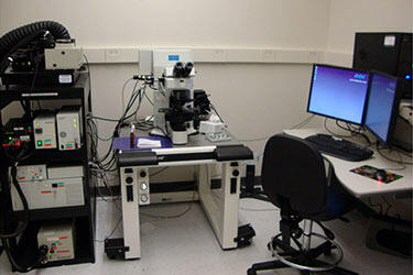
Confocal Microscopes
All microscopes are upright and all set up for DIC-IR illumination.

Olympus FV1000
Six excitation lines (405, 488, 458, 511, 568, 633), four fluorescence detection lines (two spectral units and two based on optical filters) and one detector for transmitted bright field. (Current fee $15/hour.)

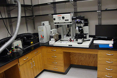
Olympus FV300
Two excitation lines (488, 568) and two detectors that can be switched for different combinations of dual fluorescence or fluorescence and transmitted light simultaneous detection. (Current fee $10/hour.)

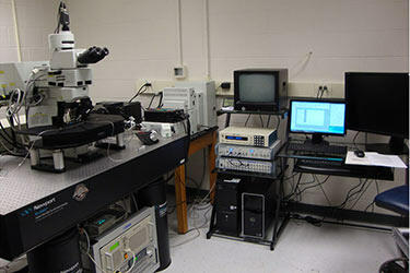
Olympus 2-photon
Two-photon excitation is obtained with a MaiTai pulsed laser (range 700-1040 nm). Single photon excitation is also possible (488, 458, 511, 559, 633 lines). Detection can be accomplished through four non-descan detectors, three confocal detectors and one transmitted light detector. (Use of this instrument and fees need to be agreed by the "2-photon user group." Current fee $20/hour.)
MCF is not responsible for any data saved on the network.

Electron Microscope
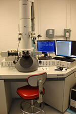
The Phillips 208S is a 100kv transmission electron microscope with excellent contrast and resolution with properly prepared specimens. The instrument, besides producing data-rich film output, is also coupled to AMT xr611 camera that permits high-resolution digital imaging.
The core provides technical help for the use of the microscope (inserting and withdrawing specimens, processing any EM negatives). Thus, usage of this instrument is always implemented with assistance by authorized personnel. The fee is $20/hour with technical assistance. EM negative usage will be charged separately depending on the level of consumption.
The core does not have enough personnel resources to help with specimen preparation.
Histocore
The Histocore is a facility designed to provide equipment support for research faculty in tissue sectioning and staining.
Equipment
- HM 550 Cryostat
- HM505 E Cryostat
- Freezing Sliding Microtome
- Vibratome
- Ultramicrotome MT 5000
- Ultramicrotome MT 6000
- Olympus Epi Fluorescence Spot Scope with RT color camera
Use of the facility will be determined by the director. All users have to be trained and approved by core personnel before being able to use equipment unsupervised. (Current fee is $10/month.)
MCF only provides equipment and training on use of the equipment. Tissue preparations are to be done by the principal investigator's lab.
Review Stations
The MCF provides three network review stations used for imaging processing and analysis:
Review I
Fluoview (FV300), Image Pro 5.1 with 3-d reconstruction, Corel Draw 12, and Office Suite
Review II
Fluoview 1.7, Image Pro 7.0, Corel Draw 12 and Office Suite
Review III (Monica)
Image Pro 5.1, Photoshop 7.0, Corel Draw 12, and Office Suite
The MCF is not responsible for any data saved on the network.
Other Hardware
Xerox Phaser 6300 Color Laser Printer
HP 1500n Monochrome Laser printer
Imacon Flextight 848 Digital negative scanner (Review III)
Policies
- Usage of the core will be determined by the director and/or the assistant directors.
- Training will be completed by the director and/or the assistant directors.
- Only authorized and trained Users are allowed to use the Microscopes in the Core Facility.
- Do not save anything on the C drive of imaging computes. This includes the desktop!
- Please save to guest or predestinated drive, you can remove your files form a designated computer in the histology core.
- All equipment must be cleaned and returned to proper positions with all personal belongings and trash removed or disposed.
Fees
|
Equipment |
Rate |
|---|---|
|
FV1000 |
$15/hour |
|
FV300 |
$10/hour |
|
2-Photon |
$20/hour |
|
Electron Microscope – TEM |
$20/hour |
|
Microscope Monthly Usage Fee |
$10/Month |
|
Histocore Monthly Usage Fee |
$10/Month |
|
Review Station Monthly Fee |
$10/Month |
|
Neurolucida/ Steroinvestigator/ Huygens |
$20/Month |
Training
-
Initial Training
Meet 1:1 with Core Personnel to gain basic understanding of theory and techniques necessary to properly operate equipment safely and effectively.
-
First Supervised Session
Learn basic troubleshooting and how to optimize your results.
-
Second Supervised Session
It’s your turn to take the lead! Expand upon your knowledge of operating your equipment of interest, while supervised by Core Personnel.
24-Hour Access
Every user has the opportunity to qualify for 24-hour access to the Microscopy Core Facility. To qualify users must:
- Log at least 20 hours of supervised usage during normal operating hours
- Demonstrate understanding of basic troubleshooting and safety procedures
- Good track record of following rules and regulations of MCF
Normal scheduling rules apply. Approval for 24-hour access is at the sole discretion of Core Personnel.
Scheduling
Please subscribe to the Wright State University Calendar system to schedule time. Time slots are 4 hour increments or less: 9 a.m.–1 p.m. | 1–5 p.m.
- A maximum of two 4-hour time slots may be reserved per week per lab.
- Only reserve time up to 3 weeks in advance
- If you are unable to use your scheduled time, please notify core personnel ASAP.
- Unscheduled time is “first come first serve”
- Delays in using the equipment over 15 minutes from scheduled time from scheduled time could result in loss of scheduled time at the discretion of Core Personnel.
PLEAE REPORT ANY PROBLEMS IMMEDIATELY TO CORE PERSONNEL.
If you have any questions or require more information about this shared instrumentation facility, please contact:
David Ladle, Ph.D.
Director, Microscopy Core Facility
259 Neuroscience Engineering Collaboration Building
937-775-4692
David.Ladle@wright.edu

